Iranian researchers develop new method to detect cancer from medical images
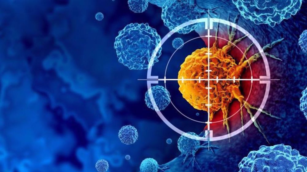
Iranian researchers have developed a new artificial intelligence-based method that improves the detection of different types of cancer from medical images, offering a promising approach for earlier diagnosis and more accurate clinical decision-making.
The study, supported by Iran's National Science Foundation (INSF), was carried out by postdoctoral researcher Yousef Sharafi at Khajeh Nasir Toosi University of Technology.
According to Sharafi, the research combines artificial intelligence, deep learning and transfer learning to extract the most important features from medical images while filtering out unnecessary data.
The approach is designed to improve the accuracy of identifying different cancers, including breast, brain and blood cancers, he added.
He explained that as cancer remains one of the world's leading causes of death, claiming more than 10 million lives each year, the development of more reliable diagnostic technologies is becoming increasingly important.
"Cancer is one of the most significant global health challenges, and advances in diagnosis and prediction can play an effective role in reducing mortality and improving treatment outcomes," Sharafi said.
🔰 Iranian researchers develop an AI-powered cancer detection method combining deep learning, transfer learning, and ensemble learning
— Iran First (@IranFirst_PTV) July 22, 2026
It improves classification accuracy for breast, brain, and blood cancers, helping earlier diagnosis and smarter medical imaging.#IranFirst pic.twitter.com/eTtxuaxn9C
The researchers first used advanced image-processing techniques and neural networks to identify and segment cancerous tumors in medical images, Sharafi said.
Once the suspicious regions were isolated, he highlighted, the system extracted key geometric, spatial and textural characteristics of cancer cells.
The extracted information was then refined using deep-learning models and optimization techniques to reduce the amount of data while preserving the most meaningful features, according to Sharafi.
Finally, he said, the images were analyzed using fuzzy neural networks and machine-learning algorithms capable of handling uncertainty, particularly in cases where the boundaries between healthy and cancerous tissue are difficult to distinguish.
The team also developed a two-level deep-learning framework specifically for analyzing breast cancer pathology and mammography images, which further improved the system's classification performance, Sharafi noted.
According to the researchers, the new method consistently outperformed conventional approaches by delivering higher accuracy in classifying cancerous tumors.
Eliminating redundant image features enabled the AI models to focus on the most informative patterns, resulting in more reliable diagnoses, Sharafi said.
"The proposed methods achieved higher accuracy and efficiency than conventional techniques in classifying cancerous tumors," Sharafi said.
"By focusing on key image features, the system improves the performance of intelligent models for cancer detection and classification."
The researchers believe the technology could contribute to the development of more advanced AI-powered medical tools for early cancer detection, disease prediction and clinical monitoring.
Sharafi also stressed that the success of AI in healthcare depends heavily on access to high-quality medical data.
"The effectiveness of artificial intelligence systems depends, above all, on standardized, accurate and well-organized medical data," he said, calling on policymakers to invest in national centers dedicated to collecting, validating and integrating cancer-related datasets.
He added that strengthening data infrastructure and training specialists in medical AI would pave the way for smarter diagnostic systems and ultimately improve healthcare outcomes for cancer patients.

Iran unveils second homegrown antivirus to counter malware and cyberattacks
Iran space agency says readying Pars 2 satellite, Martyr Soleimani constellation for launch
Iranian cardiologist wins gold medal at Swiss international invention competition


Saudi acts of aggression will not go unanswered, Yemen warns
VIDEO | News Review: Israeli aggression on Lebanon
Iran mass-producing home-grown weaponry: Air defense chief
Israeli war minister orders army to prepare for ‘all-out war’ on Palestinian Authority
Iran urges UN rights council to hold US, Israel accountable for war crimes
Report: Yemeni forces launch strikes on Saudi Aramco oil facilities in Jizan
VIDEO | The Kingdom of Compromise – II
Enemy miscalculated Iran’s ability to change war equation: Top commander


 PressTV NEWSLETTER
now
PressTV NEWSLETTER
now
 This makes it easy to access the Press TV website
